
Correlative microscopy
Correlative microscopy is an approach that benefits from imaging the same object using two different techniques.
With the increasing demand for more precise images, the pressure on the development of new technologies in microscopy is constantly growing. The term correlative microscopy refers to a group of methods that involve multiple different imaging techniques and correlate their data, leading to surprising results and scientific discoveries.
Correlation of images from two microscopes may be limited by difficult location of the region of interest or incompatibility of data acquired by different instruments under different conditions.
Correlative probe and electron microscopy
The next step of the correlative image
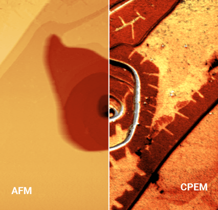
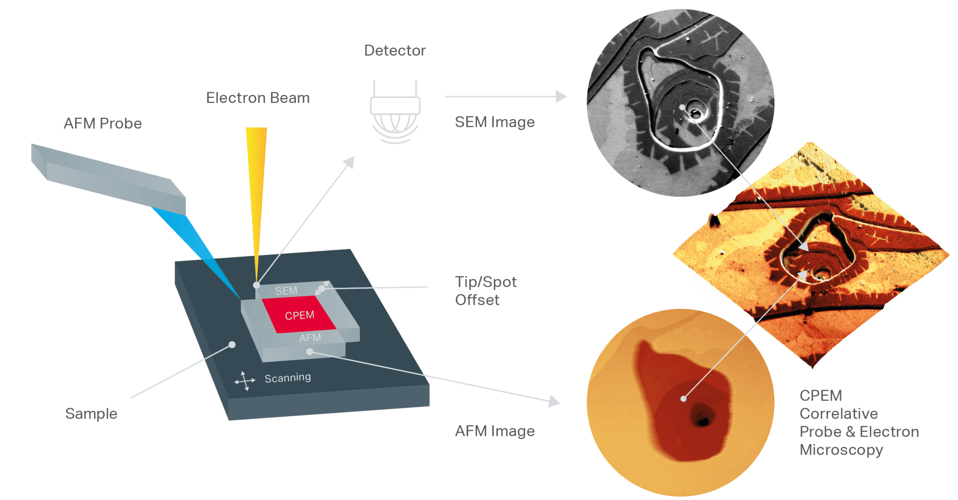
Correlative probe and electron microscopy, in short CPEM, is a novel method of multidimensional correlative imaging, allowing simultaneous acquisition of SEM and AFM data, which can be easily correlated in a 3D image. It eliminates the need for double ROI localization, enables high-precision imaging and alignment, and advanced data correlation.
Correlative Probe and Electron Microscopy (CPEM) has been developed for the application using Correlative Imaging (patented) and brings the solution, which synchronizes:
- the scanned area resolution and distortion of the image and allows to correlate AFM and SEM images acquired in real time
CPEM principle
CPEM allows the simultaneous detection and acquisition of AFM and SEM signals at the same time and in the same place.
How does it work?
During scanning, the electron beam is pointed near the tip of the AFM with a constant offset. Both remain static, while the sample is moved by the LiteScope's piezoelectric scanner. In this way, data from AFM and SEM microscopes can be acquired at the same time, in the same place and under the same conditions.
Key benefits of CPEM
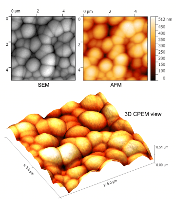
- CPEM provides multidimensional correlation images: Scanning electron microscope images are extended to 3D. With CPEM, it is possible to quickly and accurately distinguish topographical and material contrast in SEM images. CPEM correlates, appropriately, two or more SEM signals with the topography measured as SE, BSE, EBIC, etc. CPEM makes it possible to measure AFM and SEM simultaneously under the same sample conditions, at the same measurement speed, etc. The combined AFM and SEM scanning system allows a Accurate image correlation, elimination of drift and other inaccuracies.
The only true correlative microscopy
CPEM enables sample analysis in a way that was difficult or impossible with the two imaging techniques alone and opens new possibilities for advanced correlative imaging in a variety of fields such as materials science, nanotechnology, semiconductors, life sciences and many more.
LiteScope incorporates a unique imaging technique called Correlative Probe and Electron Microscopy, in short CPEM, which enables the simultaneous acquisition of AFM and SEM data. LiteScope and CPEM technology enable sample analysis in a way that was difficult or impossible with the two imaging technologies alone.
Together they provide new possibilities for advanced correlative imaging in a variety of scientific and industrial fields, such as material sciences, nanostructures, semiconductor or solar cell industry, life sciences and many more, defining their own category of correlative microscopy.
Birthday Sparks
Fashion Magazine